- PODIATRY -
FOOT OSTEOARTHRITIS

Foot osteoarthritis is a common cause of foot pain found in one in six people aged over 50, 69% of whom report disabling foot pain [1]. Osteoarthritis is the wear and tear of the cartilage within a joint which can lead to reduced range of motion and pain. The big toe joint (MTP) is the most commonly affected joint in the foot, followed by the midfoot joints (CMT, NC, TN) [1].
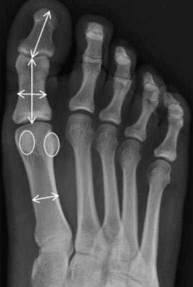
Big toe joint osteoarthritis is pain located in the 1st toe. It is found more commonly in females, with increased age and lower socioeconomic classes [1]. Structural factors associated with the condition include a flatter foot posture and longer and wider bones around the big toe joint [3, 4].
Common symptoms of big toe joint osteoarthritis include pain and stiffness within the joint [5]. Observations include a dorsal exostosis (extra growth of bone), swelling and redness [5]. Clinical assessments used to diagnose this condition include pain on palpation, limited range of motion (<64 degrees), crepitus (creaking/crunching) and a hard-end feel [5]. Big toe joint osteoarthritis can be diagnosed either clinically or with radiographs.
Mid Foot Osteoarthritis
Midfoot osteoarthritis is pain at the highlighted red regions in the above foot image. It is more prevalent in individuals aged over 75 years, in women and those in routine occupations, and is associated with obesity, pain in other weight bearing joints and previous foot and ankle injuries [7]. Structural factors associated with midfoot osteoarthritis include a flatter foot posture, greater mobility of the first metatarsal (bone attaching to the big toe joint), less range of motion in other foot joints, longer central metatarsals (long bones within the foot) and increased plantar pressures [8].
Common symptoms of midfoot osteoarthritis include tenderness across the midfoot, made worse by passively flattening the foot [10]. Having a flatter foot posture appears to be a useful predictor of symptomatic midfoot osteoarthritis [11]. Due to the complexity of the midfoot, it is inherently difficult to reliability measure the motion within these joints. Although there are measures of midfoot mobility [12], there are currently no valid clinical measures used to diagnose midfoot osteoarthritis. Therefore, currently midfoot osteoarthritis requires radiographs to confirm its diagnosis, as the current clinical assessments have either not been studied or are not strongly correlated with its diagnosis.
Treatment information
Big toe osteoarthritis
First line treatment options for foot osteoarthritis generally include NSAIDs (anti-inflammatory medication), strengthening of the foot and lower leg, footwear modifications, foot orthoses and intraarticular injections [14-17]. If conservative measures fail, surgical options are considered.
The treatment options for big toe joint osteoarthritis have only been researched in a handful of randomised controlled studies. From these studies there is some evidence for:
Sesamoid (big toe) manipulation/mobilisation, flexor hallucis longus strengthening (isometric and isotonic contractions) and gait retraining (reinforcing the functional use of the hallux flexors) [18]
Arch contouring foot orthoses [19]
Rocker-sole footwear [19]
Carbon fibre shoe stiffening inserts [20]
When looking at non randomised controlled studies, the evidence behind the treatment of big toe joint osteoarthritis suggest that people tend to respond well to conservative treatments [21, 22]. These studies found treatment options such as footwear education (wider toe box) [21], foot orthoses and corticosteroids [22] successfully reduced pain and returned patients to previous activity.
Treatment options I find useful for big toe joint osteoarthritis within the clinic include footwear education (wider toe box/rocker sole), taping, physical therapy (foot/ankle mobilisations/dry needling), custom orthotics with any modification to encourage first ray propulsion or decrease load through the forefoot. In addition, increasing strength within the lower limb to increase the foots capacity to withstand load [variations of calf raises, toe yoga (hallux/flexor hallucis brevis push downs while lifting lesser toes and vice versa] and proximal strengthening to the hip where required).
Midfoot osteoarthritis
The treatment options for midfoot osteoarthritis have only been researched in one randomised control study, which compared functional custom foot orthoses with sham orthoses [23]. This study found significant biomechanical changes and greater improvement in pain and function at 12 weeks [23]. When looking at non randomised control studies, there is evidence which found NSAIDS (anti-inflammatory medication), physical therapy, corticosteroids, foot orthoses, carbon fibre insoles and surgery beneficial in midfoot osteoarthritis [13, 24-26].
Treatment options I find useful for midfoot osteoarthritis within the clinic include footwear education (lacing techniques for the dorsal foot/encouraging less compression on the midfoot from tight footwear), custom orthotics designed to decrease the compressive forces in the midfoot and reduce plantar pressures in the heel and midfoot. As there is some evidence that people with midfoot osteoarthritis have more mobility within their first metatarsal [27], similarly to big toe joint osteoarthritis, strengthening the lower limb and muscles that insert into the foot may help stabilise the joints within the foot. In addition, people with midfoot osteoarthritis have less range of motion in the big toe and subtalar joint – so any treatments to encourage the mobility within these joints may be of benefit (foot/ankle mobilisation/dry needling/stretching/strengthening). However future research is required to further our knowledge for the best treatment options for both big toe joint and midfoot osteoarthritis, as currently the evidence is extremely limited.
Summary
Foot osteoarthritis is found in one in six people aged over 50 [1]. Both are associated with an increase in age, female sex and lower occupational classes [1, 7]. Big toe joint osteoarthritis has more clinical assessments to aid its diagnosis, compared with midfoot osteoarthritis which is harder to diagnose clinically. Evidence based treatment options are limited, however there is growing evidence behind anti-inflammatory modalities, strengthening of the foot and lower leg, footwear modifications, foot orthoses and carbon fibre shoe stiffening inserts.
If experiencing pain come in for a biomechanical assessment and be given an evidence-based treatment plan to help reduce your pain and get you back to doing what you love.
References
1. Roddy, E., et al., The population prevalence of symptomatic radiographic foot osteoarthritis in community-dwelling older adults: Cross-sectional findings from the Clinical Assessment Study of the Foot. Ann Rheum Dis, 2015. 74(1): p. 156-63.
2. Garrow, A.P., A.J. Silman, and G.J. Macfarlane, The Cheshire Foot Pain and Disability Survey: a population survey assessing prevalence and associations. Pain, 2004. 110(1-2): p. 378-384.
3. Zammit, G.V., H.B. Menz, and S.E. Munteanu, Structural factors associated with hallux limitus/rigidus: a systematic review of case control studies. journal of orthopaedic & sports physical therapy, 2009. 39(10): p. 733-742.
4. Menz, H.B., et al., Demographic and clinical factors associated with radiographic severity of first metatarsophalangeal joint osteoarthritis: cross-sectional findings from the Clinical Assessment Study of the Foot. Osteoarthritis and cartilage, 2015. 23(1): p. 77-82.
5. Zammit, G.V., S.E. Munteanu, and H.B. Menz, Development of a diagnostic rule for identifying radiographic osteoarthritis in people with first metatarsophalangeal joint pain. Osteoarthritis and cartilage, 2011. 19(8): p. 939-945.
6. Menz, H.B., et al., Radiographic classification of osteoarthritis in commonly affected joints of the foot. Osteoarthritis Cartilage, 2007. 15(11): p. 1333-1338.
7. Thomas, M.J., et al., The epidemiology of symptomatic midfoot osteoarthritis in community-dwelling older adults: Cross-sectional findings from the Clinical Assessment Study of the Foot. Arthritis Res Ther, 2015. 17(1).
8. Lithgow, M.J., et al., Foot structure and lower limb function in individuals with midfoot osteoarthritis: a systematic review. Osteoarthritis and Cartilage, 2020.
9. Menz, H.B., et al., Foot structure and function in older people with radiographic osteoarthritis of the medial midfoot. Osteoarthritis Cartilage, 2010. 18(3): p. 317-322.
10. Myerson, M., The diagnosis and treatment of injuries to the Lisfranc joint complex. The Orthopedic clinics of North America, 1989. 20(4): p. 655-664.
11. Thomas, M., et al., Clinical diagnosis of symptomatic midfoot osteoarthritis: cross-sectional findings from the Clinical Assessment Study of the Foot. Osteoarthritis Cartilage, 2015. 23(12): p. 2094-2101.
12. McPoil, T.G., et al., Arch height change during sit-to-stand: an alternative for the navicular drop test. Journal of foot and ankle research, 2008. 1(1): p. 1-11.
13. Roddy, E. and H.B. Menz, Foot osteoarthritis: latest evidence and developments. Ther Adv Musculoskelet Dis, 2018. 10(4): p. 91-103.
14. Shurnas, P.S., Hallux rigidus: etiology, biomechanics, and nonoperative treatment. Foot and ankle clinics, 2009. 14(1): p. 1-8.
15. Kurup, H. and N. Vasukutty, Midfoot arthritis-current concepts review. Journal of clinical orthopaedics and trauma, 2020. 11(3): p. 399-405.
16. Kalichman, L. and G. Hernández-Molina, Midfoot and forefoot osteoarthritis. The Foot, 2014. 24(3): p. 128-134.
17. Kunnasegaran, R. and G. Thevendran, Hallux Rigidus: Nonoperative Treatment and Orthotics. Foot and ankle clinics, 2015. 20(3): p. 401-412.
18. Shamus, J., et al., The effect of sesamoid mobilization, flexor hallucis strengthening, and gait training on reducing pain and restoring function in individuals with hallux limitus: a clinical trial. Journal of Orthopaedic & Sports Physical Therapy, 2004. 34(7): p. 368-376.
19. Menz, H.B., et al., Effectiveness of foot orthoses versus rocker‐sole footwear for first metatarsophalangeal joint osteoarthritis: randomized trial. Arthritis care & research, 2016. 68(5): p. 581-589.
20. Munteanu, S.E., et al., Shoe-stiffening inserts for first metatarsophalangeal joint osteoarthritis: a randomised trial. Osteoarthritis and Cartilage, 2021.
21. Smith, R.W., S.D. Katchis, and L.C. Ayson, Outcomes in hallux rigidus patients treated nonoperatively: a long-term follow-up study. Foot & ankle international, 2000. 21(11): p. 906-913.
22. Grady, J.F., et al., A retrospective analysis of 772 patients with hallux limitus. Journal of the American Podiatric Medical Association, 2002. 92(2): p. 102-108.
23. Halstead, J., et al., Foot orthoses in the treatment of symptomatic midfoot osteoarthritis using clinical and biomechanical outcomes: a randomised feasibility study. Clinical rheumatology, 2016. 35(4): p. 987-996.
24. Yi, T., et al., Effect of full-length carbon fiber insoles on lower limb kinetics in patients with midfoot osteoarthritis: a pilot study. American journal of physical medicine & rehabilitation, 2018. 97(3): p. 192-199.
25. Ibuki, A., et al., The effect of orthotic treatment on midfoot osteoarthritis assessed using specifically designed patient evaluation questionnaires. Prosthetics and orthotics international, 2010. 34(4): p. 461-471.
26. Rao, S., et al., Shoe inserts alter plantar loading and function in patients with midfoot arthritis. journal of orthopaedic & sports physical therapy, 2009. 39(7): p. 522-531.
27.Cowie, S., et al., Hypermobility of the first ray in patients with planovalgus feet and tarsometatarsal osteoarthritis. Foot and Ankle Surgery, 2012. 18(4): p. 237-240.
Author: Merridy Lithgow
Follow us on socials where we bring value to the run community.
![[Mo]re Than A Run Sydney 2024 🥸
So good to see so many people out running this morning and showing up (despite the rain around CP) all in aid of men’s mental health. Over 200 runners bopping around Centennial, chatting and starting conversati](https://images.squarespace-cdn.com/content/v1/5a77ea35dc2b4a0bdb3fc4b9/1732941224838-WML7GNF86XR3TXA8ZQ5L/image-asset.jpeg)